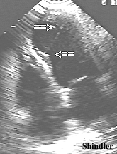
Left ventricular false tendons are fibrous or fibromuscular bands that stretch across the left ventricle from the septum to the free wall. They can also tether to a papillary muscle, but unlike the chordae tendineae, do not connect to the mitral leaflets. They are anatomic variants that should not be mistaken for abnormalities such as tumors, subaortic membranes, thrombus borders, or septal hypertrophy. They have been noted in patients with childhood murmurs and in patients with arrhythmias.
References
Badano L, Piazza R, Bisignani G, Nicolosi GL.
Echocardiographic features of left ventricular aneurysm and
false tendon in a patient with postinfarction pseudoaneurysm
after aneurysmectomy.
G Ital Cardiol 1993 Mar;23(3):295-9
Three years after the repair of a true left ventricular aneurysm, a 62-year-old man was admitted with angina and heart failure. The two-dimensional echocardiogram revealed a uniformly dilated left ventricle with a large apical aneurysm, in which a thin, continuous, band-like echogenic structure, extending from the interventricular septum to the antero-lateral wall could be visualized. That structure was initially interpreted as a left ventricular false tendon. Color Doppler flow imaging, however, showed a continuous, phasic flow crossing the band-like structure. Thus, the diagnosis of a huge apical pseudoaneurysm was made and subsequently confirmed by angiographic findings.
Calabro MP, De Luca F, Consolo S, Falcone G, Oreto G.
Left ventricular false tendon: the most frequent cause of innocent
murmur in childhood?
G Ital Cardiol 1992 Jan;22(1):19-24
To assess the incidence of false tendons in children with a murmur classified as innocent - two groups of subjects were selected. Group A consisted of 253 children with: 1) systolic ejection murmur; 2) normal electrocardiogram and 3) absence of clinical data suggesting cardiac disease. Group B consisted of 240 children clinically free of cardiac disease, and without any cardiac murmur. One hundred and sixty-one children of group A (63.6%) had a left ventricular false tendon. Three out of the 161 had a small ventricular septal defect (the false tendon was the only abnormal finding in 158 of the children). Seventy one (28.1%) of group A had an normal echocardiogram; 21 (8.3%) had congenital heart disease. In group B, only 33 subjects (13.8%) had a false tendon. The different incidence of false tendons in the two groups (63.6% versus 13.8%) was statistically significant (p less than 0.01).
Goicolea FJ et al.
False tendon rupture simulating chordal
rupture after percutaneous mitral balloon dilation.
A report of two cases.
Eur Heart J 1991 Jul;12(7):829-31.
Two cases of ruptured false tendons followed percutaneous balloon dilation of the mitral valve in one case, and combined mitral and aortic balloon valvotomy in the other. No adverse effects were noted following this complication.
Morin LR et al.
Prenatal diagnosis of left ventricular false
tendon.
Acta Obstet Gynecol Scand 1989;68(5):463-4
Kudoh Y, Hiraga Y, Iimura O.
Benign ventricular tachycardia in
systemic sarcoidosis--a case of false tendon.
Jpn Circ J 1988 Apr;52(4):385-9.
A 22-year-old female patient was diagnosed as having systemic sarcoidosis with pulmonary, skin and ocular lesions, and ventricular tachycardia in resting ECG. Although cardiac sarcoidosis was strongly suspected at diagnosis, no clinical symptom such as palpitation or syncope developed during the three year observation period. Cardiac silhouette was unchanged in chest X-ray and 201thallium myocardial scintigraphy revealed no abnormality. Ventricular complex was suppressed by exercise or tachycardia. Two-dimensional echocardiogram showed abnormal fascicular bands attached from the mid-septum to the apex (false tendon). Therefore, it was concluded that this benign form of ventricular tachycardia might be due to the false tendon, rather than to the cardiac involvement of sarcoidosis. The cause of arrhythmia is important when evaluating the prognosis of a patient with a systemic disease.
Okamoto M et al.
Visualization of the false tendon in the left
ventricle with echocardiography and its clinical significance.
J Cardiogr 1981 Mar;11(1):265-70.
False tendons were best detected in the apical long axis view in 61 of 132 consecutive patients. The incidence did not seem to be related to the underlying conditions. The false tendon was usually a string, a few millimeters in width, crossing the ventricular cavity from the vicinity of the papillary muscles to the interventricular septum. In a few patients it looked as Y-figure and net like. Sometimes, several sticks of the false tendon were detected. It was observed to be stretched in diastole and relaxed in systole. On the M-mode echocardiogram the false tendon was displayed as a linear echo moving with heart beat. False tendons seen near the interventricular septum, exhibited a motion so similar to that of the interventricular septum that they should be carefully differentiated from the echo of the left ventricular surface of the septum. In 2 patients with valvular heart disease, the false tendon was observed to be fluttering in diastole. Echocardiography was more useful in detecting false tendons than left ventriculography.
Back to Hand-Held Echocardiography Home Page.
 The contents and links on this page were last verified by Dr. Olga Shindler on February 7, 2005.